
Presenting Case: The Cat with One Kidney Left
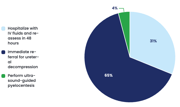
This week’s case showed strong alignment between practicing veterinarians and our specialists, with 65% of responding DVMs selecting immediate referral for ureteral decompression as their next step. Our specialists agreed, emphasizing that when ureteral obstruction threatens a cat’s remaining functional kidney, time matters. Fluids may support the patient, but they cannot resolve a mechanical obstruction—and delaying decompression can increase the risk of irreversible renal damage.
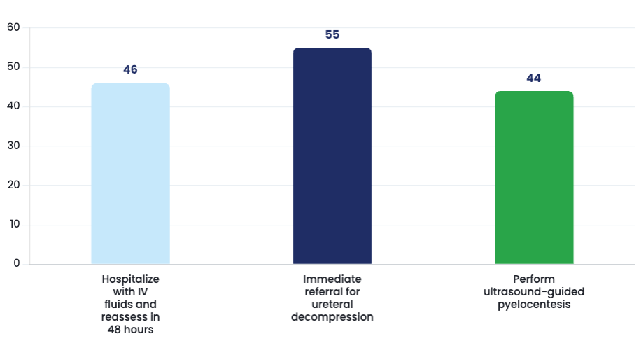
Preferred Treatment by Responding DVMs Effectiveness of Treatments
Patient Recap
9-year-old Spayed Female Domestic Shorthair
Presenting Complaint
Acute anorexia and vomiting for two days.
History
The owner reports that “Luna” stopped eating approximately 48 hours ago. She has vomited three times over the past day and has become increasingly withdrawn. She has spent most of the last 24 hours hiding under the bed and has shown little interest in food or water.
Additional history includes:
- Indoor-only cat
- Diagnosed with IRIS Stage II chronic kidney disease one year ago
- Receiving a prescription renal diet
- No history of urinary tract obstruction
- No toxin exposure
- No current medications besides omega-3 supplementation
Physical Examination
- Quiet but responsive
- Body Condition Score: 4/9
- Temperature: 100.1°F
- Heart Rate: 168 bpm
- Respiratory Rate: 32 bpm
- Approximately 7% dehydrated
- Moderate pain on cranial abdominal palpation
- Left kidney palpates small and irregular
- Right kidney enlarged and painful
- Mild halitosis consistent with uremia
Diagnostics
CBC
- Mild non-regenerative anemia (PCV 27%)
- White blood cell count within normal limits
Chemistry Panel
- Creatinine: 4.2 mg/dL
- BUN: 82 mg/dL
- Phosphorus: 7.8 mg/dL
- Potassium: 5.9 mmol/L
- SDMA: Elevated
Urinalysis
- USG: 1.020
- No bacteria identified
- No significant sediment
Abdominal Ultrasound
- Left kidney small and irregular with chronic architectural changes
- Right renal pelvis moderately dilated
- Proximal right ureter contains a 3 mm mineralized structure
- Mild perirenal fluid present
Specialist Analysis
How a General Practitioner Might Approach This Case
Cats with acute kidney injury often present with similar clinical signs.
Vomiting. Anorexia. Dehydration. Azotemia.
Because intravenous fluid therapy remains the cornerstone of treatment for many renal diseases, it is understandable that many clinicians initially recommend hospitalization with aggressive fluid therapy before considering referral.
After all, many cats improve significantly once dehydration is corrected. However, specialists recognize that not all acute kidney injuries are medical diseases. Some are mechanical emergencies. This case represents one of those situations.
How Specialists Typically View This Case
From an internal medicine and interventional radiology perspective, the most appropriate recommendation is:
Immediate Referral for Ureteral Decompression
The ultrasound findings completely change the clinical picture. Rather than progressive chronic kidney disease alone, Luna has developed a ureteral obstruction affecting her only remaining functional kidney.
This is no longer simply an azotemic cat. It is a cat with a kidney that cannot drain.
Why One Obstructed Kidney Can Become an Emergency
At first glance, unilateral ureteral obstruction may not seem immediately life-threatening. After all, the opposite kidney should continue functioning. Except in this patient…
The left kidney is already severely compromised by chronic kidney disease. Functionally, Luna has been relying almost entirely on her right kidney. Once the right ureter became obstructed, her remaining renal function rapidly declined. Within hours, pressure began building inside the renal pelvis. As pressure increases, glomerular filtration decreases.
Blood flow to the kidney falls. Without intervention, irreversible nephron damage can begin within days. The concern is no longer simply elevated creatinine. It is permanent loss of the patient’s remaining renal function.
Why IV Fluids Alone Are Not Enough
Fluid therapy is critically important. These patients are almost always dehydrated and uremic. However, fluids cannot remove an obstruction. No matter how well hydrated the patient becomes, urine cannot bypass a mineralized ureterolith. Continuing to increase fluid administration may actually worsen renal pelvic pressure while providing little improvement in kidney function. Supportive care should accompany definitive treatment—not replace it.
Why Immediate Referral Is Recommended
Over the past decade, treatment of feline ureteral obstruction has changed dramatically. Historically, ureterotomy carried significant complications and variable success.
Today, most referral centers perform either:
- Subcutaneous ureteral bypass (SUB) placement
- Ureteral stent placement
Both procedures restore urine flow while preserving functional renal tissue. Multiple studies have demonstrated substantially improved survival and long-term renal outcomes when obstruction is relieved promptly.
Timing matters. The longer obstruction persists, the lower the likelihood of meaningful renal recovery.
Why Not Start Antibiotics?
There is no evidence of bacterial infection. The urinalysis is unremarkable. No pyuria or bacteriuria is present. While pyelonephritis certainly remains a differential diagnosis in cats with renal disease, nothing in this case suggests infection as the primary cause. Empirical antibiotics delay appropriate treatment without addressing the obstruction.
Why Not Perform a Pyelocentesis?
Ultrasound-guided renal pelvic aspiration can occasionally be useful for obtaining sterile urine samples when pyelonephritis is suspected. However, this patient has no evidence of infection. Furthermore, pyelocentesis does nothing to relieve the obstruction itself. It may provide additional diagnostic information, but it does not alter the underlying disease process. Specialists reserve this procedure for carefully selected cases rather than routine management of ureterolithiasis.
The Biggest Teaching Point
One of the most important lessons in feline nephrology is that creatinine does not tell the entire story. Many clinicians understandably focus on the number. Specialists focus on why the number increased. This cat’s worsening azotemia was not simply progression of chronic kidney disease. It represented an acute obstruction superimposed on chronic renal insufficiency. Recognizing that distinction changes everything. One disease is managed medically. The other requires intervention.
Practical Application in General Practice
When evaluating cats with acute azotemia, specialists encourage clinicians to ask four questions.
Could this be obstructive? Always evaluate both kidneys and both ureters.
Are both kidneys functioning? A unilateral obstruction becomes much more serious when the opposite kidney is already compromised.
Does ultrasound demonstrate renal pelvic dilation? Hydronephrosis should always raise concern for ureteral obstruction.
Will medical therapy correct the underlying problem? If the answer is no, referral should be discussed early.
Specialist Takeaway
Most cats with chronic kidney disease experience gradual progression over months or years. This patient changed dramatically over just two days. That rapid deterioration was the clue.
Rather than worsening chronic kidney disease alone, Luna developed an obstructive ureterolith affecting her only functional kidney. While intravenous fluids remain an important component of stabilization, they cannot relieve a mechanical obstruction. For this reason, our internal medicine specialist recommends:
Immediate Referral for Ureteral Decompression
Whether accomplished through a subcutaneous ureteral bypass (SUB) device or ureteral stent placement, restoring urine flow offers the best opportunity to preserve remaining renal function and improve long-term outcome.
The broader lesson applies well beyond feline nephrology.
Whenever a patient with chronic disease suddenly worsens, clinicians should ask whether they are seeing progression of the original condition—or the development of an entirely new problem.
The answer often determines whether the patient needs supportive care or immediate intervention.
References (MLA)
Berent, Allyson C., and Chick Weisse. “Management of Benign Ureteral Obstruction in Cats.” Journal of Feline Medicine and Surgery, vol. 16, no. 1, 2014, pp. 33–39.
Kyles, Ashley E., et al. “Clinical, Clinicopathologic, Radiographic, and Ultrasonographic Abnormalities in Cats with Ureteral Calculi.” Journal of the American Veterinary Medical Association, vol. 226, no. 6, 2005, pp. 932–936.
Berent, Allyson C. “Subcutaneous Ureteral Bypass Devices in Cats with Benign Ureteral Obstruction.” Veterinary Clinics of North America: Small Animal Practice, vol. 50, no. 2, 2020, pp. 305–324.
IRIS (International Renal Interest Society). IRIS Guidelines for the Diagnosis and Management of Acute Kidney Injury and Chronic Kidney Disease in Cats, 2023.