

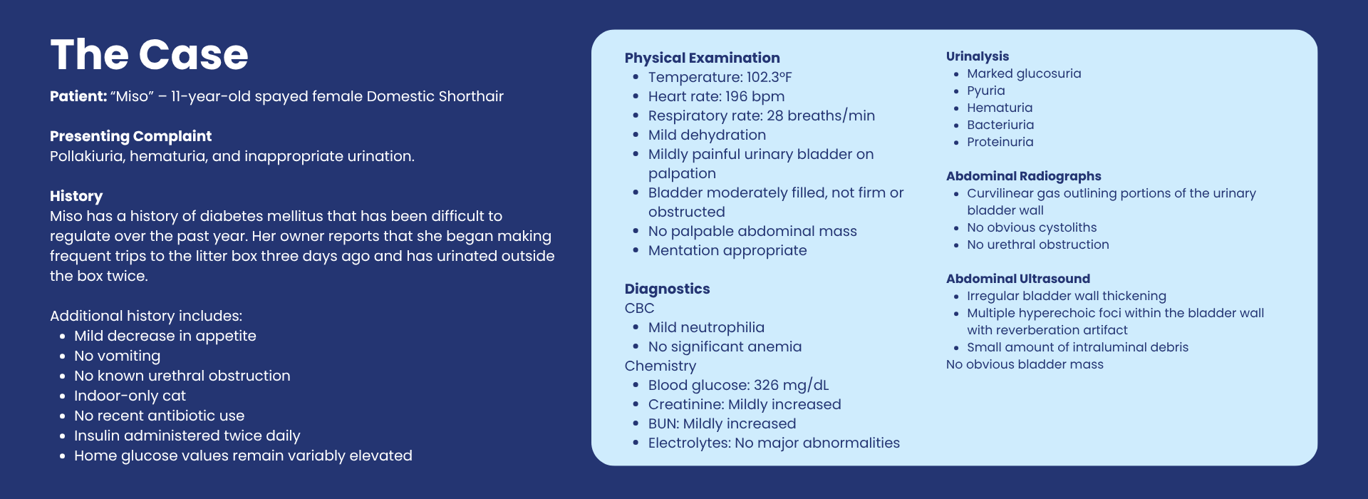
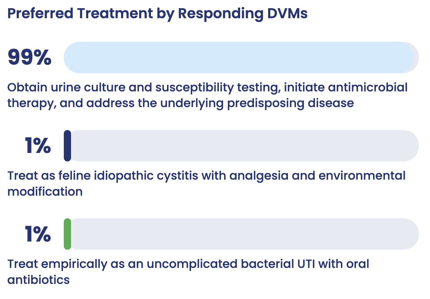
99% of responding DVMs selected the same approach recommended by specialists. In a case where the presenting signs could easily point toward a routine urinary tract infection, veterinarians overwhelmingly recognized that the presence of gas within the bladder wall changed the clinical picture. The near-unanimous response shows strong alignment around the need for urine culture and susceptibility testing, appropriate antimicrobial therapy, and management of the underlying predisposing disease.
When the Imaging Changes the Case
At first glance, Miso’s presentation looks familiar.
Pollakiuria. Hematuria. Urinating outside the litter box. Pyuria and bacteriuria on urinalysis.
In everyday general practice, those findings could reasonably lead a clinician toward one of the most common explanations: bacterial cystitis, followed by antimicrobial treatment.
But this case included one finding that made it much harder to treat as routine:
Gas within the bladder wall.
And judging by the survey results, responding veterinarians recognized its significance.
Rather than seeing the usual split between a practical general-practice approach and the more diagnostic-heavy approach favored by specialists, nearly all responding DVMs selected the same answer recommended by the specialists: obtain urine culture and susceptibility testing, initiate appropriate antimicrobial therapy, and address the underlying predisposing disease.
That degree of agreement is notable because the case begins with signs that are easy to categorize as a familiar lower urinary tract problem. The difference was recognizing when one abnormal finding changed the clinical picture.
The Finding That Changes the Differential
Gas within the urinary bladder wall is not a typical feature of uncomplicated bacterial cystitis.
It raises concern for emphysematous cystitis, an uncommon infection characterized by gas within the bladder wall and/or lumen produced by gas-forming microorganisms. Diagnostic imaging is central to recognizing the condition, with radiography and ultrasonography commonly used to identify intramural or intraluminal gas.
Reported organisms include Escherichia coli, Klebsiella spp. and other gas-producing bacteria. In a veterinary retrospective study of 38 animals with emphysematous cystitis, including two cats, 92.1% had a positive urine culture, and E. coli was the most frequently identified organism.
So while Miso’s urinary signs may look routine, the imaging makes the case anything but routine.
Why Diabetes Matters
Miso’s diabetes is also more than a background diagnosis.
Diabetes mellitus has long been associated with an increased risk of bacterial urinary tract infection, and diabetic patients are frequently represented in reports of emphysematous urinary infections.
In one study of 141 diabetic cats, 18 had a positive urine culture, with E. coli accounting for 67% of isolates.
Glucosuria may provide substrate that can support bacterial fermentation, while concurrent metabolic disease can alter host defenses and create conditions that make infection more difficult to resolve.
That leads to two interconnected clinical objectives:
- Identify and appropriately treat the infection.
- Address the condition that may be contributing to its development or persistence.
Treating only the bladder infection without considering diabetic control risks addressing only part of the problem.
Why Culture Matters Here
This is where the specialist recommendation becomes particularly important.
For a straightforward episode of lower urinary tract disease, clinicians may sometimes begin treatment based on the clinical picture. But this patient has several features that move the case away from a simple presentation: a significant comorbidity, bacteriuria and an unusual imaging abnormality.
Current ISCAID guidance emphasizes bacterial culture and susceptibility testing when managing more complicated or recurrent urinary infections and recommends investigating underlying factors that could contribute to persistence or recurrence.
A culture can:
- Confirm the presence of bacterial infection.
- Identify the causative organism.
- Guide antimicrobial selection.
- Identify resistance.
- Support more deliberate antimicrobial stewardship.
If Miso requires immediate treatment, antimicrobial therapy may still begin while culture results are pending. The important point is that the urine sample should ideally be obtained first so treatment can ultimately be directed by the organism and susceptibility results.
Why Not Simply Treat It as an Uncomplicated UTI?
Because the imaging has already told us that the disease is behaving differently.
Intramural gas suggests a gas-forming infectious process rather than routine cystitis. Treating empirically without investigating further creates the possibility of choosing an ineffective antimicrobial, overlooking resistance, or failing to address an underlying predisposition.
Perhaps more importantly, it risks ignoring the finding that makes this case clinically distinctive.
The lesson is not that empiric treatment is always inappropriate.
It is that once the case stops looking uncomplicated, the diagnostic strategy should change with it.
Why Not Go Straight to Surgery?
The opposite reaction is also possible.
Gas in the bladder wall looks dramatic, and emphysematous cystitis sounds like a disease that should require aggressive surgical intervention.
But surgery is not automatically indicated.
Published veterinary and human literature describes medical management as the primary approach in many patients, combining appropriate antimicrobial therapy with management of predisposing disease. Surgical intervention is generally reserved for complications such as necrosis, perforation, obstruction, structural abnormalities or failure of medical management.
Nothing in Miso’s presentation suggests that immediate cystotomy should be the first step.
Why Not Feline Idiopathic Cystitis?
Feline idiopathic cystitis remains an important differential for cats presenting with lower urinary tract signs.
But it is not a diagnosis that overrides evidence of bacterial disease.
In this case, pyuria, bacteriuria and gas within the bladder wall collectively shift the clinical reasoning toward infection. Analgesia and environmental management may still have roles in the patient’s broader care, but they would not address the primary infectious process identified here.
What This Survey Result Tells Us
This is where the response pattern becomes particularly interesting.
In many Clinical Outcomes cases, there is a visible difference between the first step selected by general practitioners and the recommendation favored by specialists. Those differences often reflect the realities of general practice: balancing diagnostic certainty, cost, availability, clinical experience and the need to make decisions quickly.
That divide largely disappeared here.
Nearly all responding veterinarians recognized that this was not simply another cat with lower urinary tract signs.
They identified the imaging abnormality, connected it with the patient’s underlying diabetes, and selected the more comprehensive diagnostic and treatment strategy recommended by the specialists.
That suggests an important point about clinical decision-making:
Common presentations do not necessarily lead to routine decisions when the case provides a strong enough signal to reconsider the diagnosis.
For Miso, that signal was gas within the bladder wall.
The urinary signs were familiar.
The bacteriuria was familiar.
The diabetic patient was familiar.
But one abnormal imaging finding reframed all of them.
And in this case, general practitioners and specialists overwhelmingly arrived at the same place.
Specialist Takeaway
The recommended approach is to obtain a urine culture and susceptibility test, institute appropriate antimicrobial therapy, and address the underlying predisposing disease.
The broader lesson may be even more useful:
When a common presentation contains an uncommon finding, that finding deserves disproportionate attention.
Sometimes the most important part of a case is not the symptom seen most often.
It is the detail that does not belong.
References
- Moon R, Biller DS, Smee NM. Emphysematous cystitis and pyelonephritis in a nondiabetic dog and a diabetic cat. J Am Anim Hosp Assoc. 2014;50(2):124-129. doi:10.5326/JAAHA-MS-5972.
- Evangelista LSM, et al. Emphysematous cystitis: review of current literature, diagnosis and management challenges. Vet Med (Auckl). 2019.
- Mannucci T, et al. Emphysematous cystitis: retrospective evaluation of predisposing factors and ultrasound features in 36 dogs and 2 cats. Vet Radiol Ultrasound. 2019.
- Bailiff NL, Nelson RW, Feldman EC, et al. Frequency and risk factors for urinary tract infection in cats with diabetes mellitus. J Vet Intern Med. 2006;20(4):850-855.
- Weese JS, Blondeau J, Boothe D, et al. International Society for Companion Animal Infectious Diseases (ISCAID) guidelines for the diagnosis and management of bacterial urinary tract infections in dogs and cats. Vet J. 2019;247:8-25. doi:10.1016/j.tvjl.2019.02.008.