

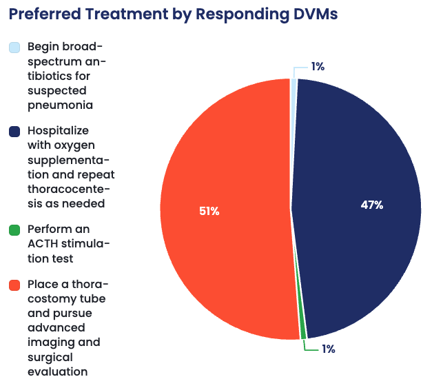
More than half of responding DVMs landed on the same next step as the specialist. In this case of acute, nontraumatic bilateral pneumothorax, 51% selected placement of a thoracostomy tube followed by advanced imaging and surgical evaluation as their recommended next step.
Another 47% chose continued hospitalization with oxygen supplementation and repeat thoracocentesis as needed, showing just how close the decision was between continued stabilization and moving toward more definitive intervention.
So what makes thoracostomy tube placement and surgical evaluation the preferred next step in this case? Let’s take a closer look at the specialist’s reasoning.
How a General Practitioner Might Approach This Case
This case presents an interesting decision point because the immediate treatment has worked.
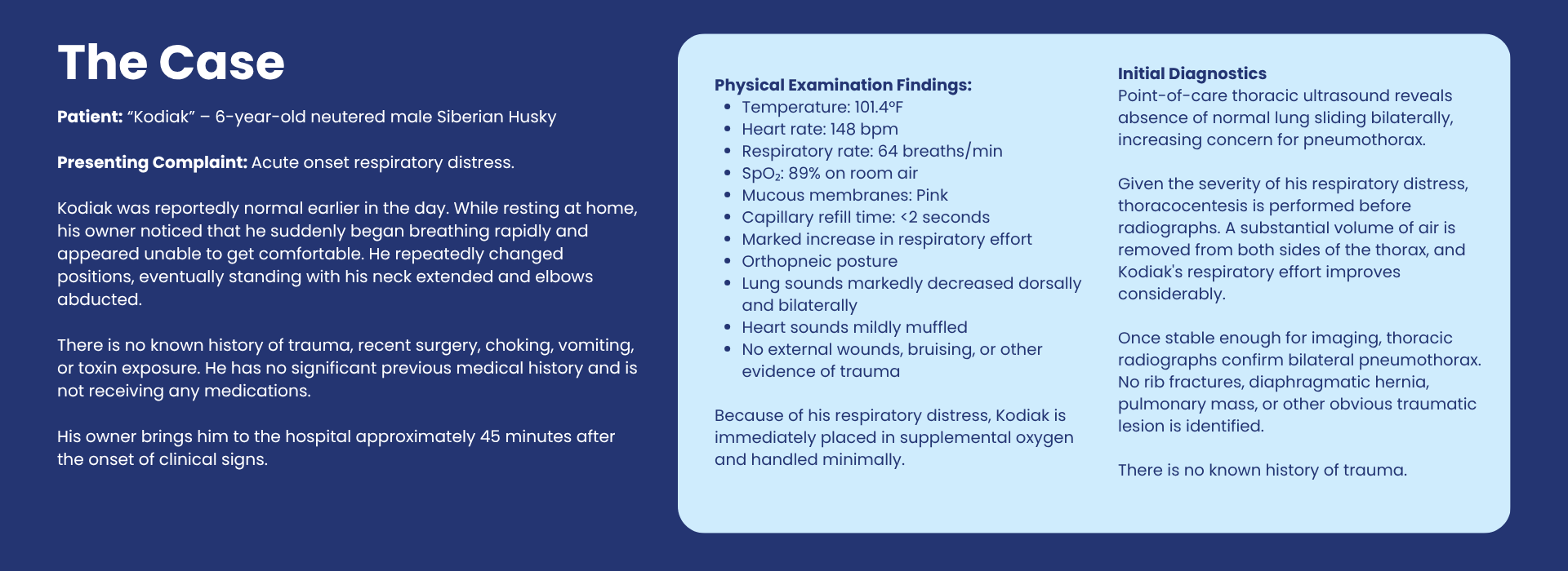
Kodiak arrived in significant respiratory distress. Pneumothorax was recognized quickly, thoracocentesis was performed, and his breathing improved.
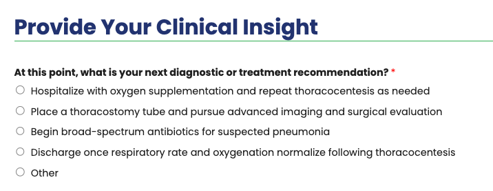
From a general practice perspective, Answer A may therefore feel like a reasonable next step: hospitalize, provide oxygen, closely monitor respiratory effort, and repeat thoracocentesis if air begins accumulating again.
In some forms of pneumothorax, particularly when the underlying insult is self-limiting, conservative management may be successful.
The absence of an obvious pulmonary lesion on radiographs may also provide some reassurance.
But there is one major problem:
We still don’t know why air entered Kodiak’s pleural space in the first place.
There was no trauma. No recent procedure. No obvious external injury.
That changes the clinical significance of the pneumothorax.
How Specialists Typically View This Case
A surgeon or emergency and critical care specialist would likely approach Kodiak’s case as spontaneous pneumothorax until proven otherwise.
And in dogs, spontaneous pneumothorax should raise immediate concern for an underlying pulmonary lesion—particularly a ruptured pulmonary bulla or bleb. These lesions are among the most common causes of spontaneous pneumothorax in otherwise healthy dogs.
Kodiak’s signalment adds to that suspicion.
Spontaneous pneumothorax is seen disproportionately in large, deep-chested dogs, and Siberian Huskies and other Northern breeds are overrepresented.
The fact that thoracocentesis improved his respiratory distress is important—but it doesn’t mean the underlying problem has resolved.
It means the consequence of the lesion has temporarily been corrected.
For this reason, the specialist answer is:
- Place a thoracostomy tube and pursue advanced imaging and surgical evaluation.
What Is Happening in Spontaneous Pneumothorax?
Under normal circumstances, negative pressure within the pleural space keeps the lungs expanded against the thoracic wall.
When air enters that space, negative intrathoracic pressure is lost.
As pleural air accumulates, the lungs cannot fully expand, resulting in:
- Reduced functional lung volume
- Impaired ventilation
- Ventilation-perfusion mismatch
- Hypoxemia
- Increasing respiratory effort
If enough air accumulates—or continues accumulating faster than it can be removed—the condition can become life-threatening.
In primary spontaneous pneumothorax, the underlying problem is frequently rupture of an abnormal air-filled structure within the lung.
Bullae vs. Blebs
A bleb is a small collection of air associated with the visceral pleural surface, while a bulla represents a larger air-filled space within abnormal pulmonary tissue.
When one ruptures, air escapes from the respiratory tract into the pleural space.
That leak may temporarily seal.
Or it may continue.
And that uncertainty is precisely why a patient can look significantly better after thoracocentesis but deteriorate again hours later.
Why Normal Radiographs Don’t End the Investigation
This is one of the most important lessons from Kodiak’s case. Thoracic radiographs are excellent for answering:
“Is there a pneumothorax?”
They are much less reliable for answering:
“What caused the pneumothorax?”
In one canine study, radiographs detected pneumothorax in all 12 affected dogs but identified the underlying bullae or blebs in only 2 of 12 dogs. CT identified bullae or blebs in 9 of 12 and identified substantially more affected lung lobes than radiography.
Another series of dogs with surgically confirmed pulmonary blebs and bullae found radiographic evidence of those lesions in only one of 12 dogs.
So Kodiak’s radiographs showing “no obvious pulmonary lesion” should not be interpreted as:
There is no pulmonary lesion.
Instead, the more appropriate interpretation is:
The cause is not apparent on radiographs.
That distinction matters.
Why CT Becomes Valuable
Once the patient has been stabilized, thoracic CT can help identify pulmonary bullae, blebs, masses, areas of diseased lung, or other abnormalities that may be responsible for the air leak.
It can also help surgeons understand whether abnormalities appear focal, multifocal, or bilateral and assist with procedural planning. CT is considerably more sensitive than conventional radiography for identifying bullae and blebs associated with spontaneous pneumothorax.
But CT is not necessarily the end of the diagnostic process.
Small lesions can still be missed, and surgical exploration may ultimately be required to identify and remove the source of leakage.
Why Place a Thoracostomy Tube?
Thoracocentesis is an excellent emergency stabilization procedure.
It removes accumulated pleural air and allows the lungs to re-expand.
But if air continues leaking, repeatedly inserting a needle into the thorax is not an ideal long-term management strategy.
A thoracostomy tube provides ongoing access to the pleural space and permits intermittent or continuous evacuation of air while the patient is stabilized and the underlying cause is investigated.
The important distinction is:
The chest tube manages the pneumothorax. It does not necessarily treat its cause.
If a ruptured pulmonary bulla continues leaking, definitive treatment may require surgical resection of the affected lung tissue.
Why Surgery Enters the Conversation Early
This is where the specialist perspective may differ most significantly from a “wait and see” approach.
Historically, spontaneous pneumothorax has been treated both medically and surgically.
But available canine outcome data generally favor surgical treatment when pulmonary bullae or blebs are responsible.
A retrospective study of 64 dogs found significantly lower recurrence and mortality among surgically managed dogs compared with dogs receiving nonsurgical management alone. The authors recommended early surgical intervention for definitive diagnosis and treatment.
More contemporary surgical series also demonstrate favorable long-term outcomes. In one multicenter study, surgically treated dogs had reported two- and five-year survival rates of 88.4% and 83.5%, respectively.
A separate study of 110 surgically managed dogs found that lung lobectomy resolved pneumothorax in most patients; when recurrence occurred, it was concentrated primarily in the early postoperative period.
That does not mean every dog with spontaneous pneumothorax should be rushed directly from thoracocentesis into surgery.
Stabilization comes first.
But once a spontaneous pneumothorax is identified, particularly in a patient whose signalment is highly suggestive of bullous disease, early referral and surgical evaluation become an important part of the treatment plan.
Why Not Answer A: Hospitalize and Repeat Thoracocentesis?
This is probably the most tempting alternative.
And it isn’t unreasonable as an initial stabilization strategy.
Kodiak absolutely should be hospitalized and monitored.
The problem is treating repeated thoracocentesis as the entire plan.
If pleural air repeatedly accumulates, something is continuing to leak. Repeated drainage treats that consequence without identifying or correcting its source.
Historical outcome data also suggest substantially greater recurrence with nonsurgical management of canine spontaneous pneumothorax.
For a stable patient with a very small pneumothorax or a clearly identified self-limiting cause, conservative management may sometimes be appropriate.
But Kodiak has already presented with significant bilateral pneumothorax and hypoxemia, without an identifiable traumatic cause.
That warrants a more aggressive investigation.
Why Not Answer C: Begin Broad-Spectrum Antibiotics?
Pneumonia, pulmonary abscessation, migrating foreign bodies, fungal disease, neoplasia, heartworm disease, and other pulmonary conditions can occasionally cause secondary spontaneous pneumothorax.
But nothing in Kodiak’s current presentation specifically supports bacterial pneumonia.
There is no reported fever, productive cough, radiographic pulmonary infiltrate, or other compelling evidence of bacterial infection.
Starting empirical antibiotics does not address the immediate pleural air leak and could distract from identifying its actual source.
If subsequent imaging, cytology, culture, or surgical findings indicate infection, antimicrobial therapy can then be selected appropriately.
Why Not Answer D: Discharge After Successful Thoracocentesis?
This is the riskiest option.
Clinical improvement following thoracocentesis tells us that removing pleural air improved pulmonary expansion.
It tells us nothing about whether the leak has stopped.
A patient with a persistent or intermittent pulmonary leak can rapidly accumulate pleural air again and return to respiratory distress.
Kodiak therefore requires hospitalization and monitoring even if he appears dramatically better immediately after thoracocentesis.
Feeling better after drainage is not the same as being cured.
Practical Application in General Practice
The first priority in any dyspneic patient is stabilization—not obtaining the perfect set of radiographs.
When pneumothorax is strongly suspected in a patient experiencing substantial respiratory compromise, therapeutic thoracocentesis may appropriately precede extensive diagnostic imaging.
Once the patient is stabilized, ask a second question:
Why did this pneumothorax happen?
A history of trauma provides one pathway.
But when there is no trauma or iatrogenic explanation, spontaneous pneumothorax should move much higher on the differential list.
And in a large, deep-chested dog—particularly a Siberian Husky or another predisposed breed—the possibility of pulmonary bullae or blebs deserves serious consideration.
That is the point at which referral can change the outcome.
Specialist Takeaway
Thoracocentesis can save the patient’s life, but it may only be treating the symptom.
In a dog presenting with spontaneous pneumothorax, particularly without trauma and with a signalment associated with pulmonary bullae or blebs, specialists recommend looking beyond the immediate improvement following pleural air evacuation.
Thoracostomy tube placement can provide continued control of pleural air while CT and surgical evaluation identify the source of the leak and determine whether definitive surgical treatment is warranted.
The key clinical lesson is simple:
When air appears in the pleural space without an obvious reason, don’t stop once the patient starts breathing better. Find out why it happened.
References
American College of Veterinary Surgeons. “Spontaneous Pneumothorax.” ACVS, American College of Veterinary Surgeons.
Lipscomb, Victoria J., Robert J. Hardie, and Richard R. Dubielzig. “Spontaneous Pneumothorax Caused by Pulmonary Blebs and Bullae in 12 Dogs.” Journal of the American Animal Hospital Association, vol. 39, no. 5, 2003, pp. 435–445.
Puerto, David A., et al. “Surgical and Nonsurgical Management of and Selected Risk Factors for Spontaneous Pneumothorax in Dogs: 64 Cases (1986–1999).” Journal of the American Veterinary Medical Association, vol. 220, 2002, pp. 1670–1674.
Au, J. J., et al. “Use of Computed Tomography for Evaluation of Lung Lesions Associated with Spontaneous Pneumothorax in Dogs: 12 Cases (1999–2002).” Journal of the American Veterinary Medical Association, vol. 228, 2006, pp. 733–737.
This one should give you a very good survey split between A and B. A feels clinically defensible because the patient improves after thoracocentesis, while B captures the specialist concern that a spontaneous pneumothorax in this signalment often requires investigation and definitive management rather than repeated drainage alone.