

This is a network committed to advancing clinical understanding through real-world outcomes. We believe veterinarians hold unparalleled insight into what truly works in practice. Each week, we pose one focused clinical question to the veterinary community. On Mondays, we present the question. On Fridays, we share your responses—highlighting the treatments and protocols delivering the best outcomes. Together, we’re building a living library of frontline veterinary wisdom.
CLINICAL PERSPECTIVE
This Week’s Sticky Case
Patient
7-year-old Female Spayed Golden Retriever
Presenting Complaint
Progressive weight loss and intermittent diarrhea
History
Owner reports:
- Soft stools for approximately 4 months
- Episodes of diarrhea occurring every 1–2 weeks
- Appetite remains excellent
- Approximately 8-pound weight loss over the past 3 months
- No vomiting
- No travel history
- Monthly heartworm/flea prevention current
- Previously completed:
- Metronidazole trial
- Probiotic supplementation
- OTC sensitive stomach diet
Minimal improvement reported.
Physical Examination
- Bright and alert
- BCS 4/9
- Mild muscle loss over epaxial muscles
- Mildly thickened intestinal loops on abdominal palpation
- No peripheral lymphadenopathy
- Temperature: 101.8°F
- HR: 90 bpm
Diagnostics
CBC
- Mild mature lymphocytosis
- Mild eosinophilia
Chemistry
- Albumin: 2.2 g/dL (Low)
- Globulin: Normal
- Cholesterol: Low
- ALT: Normal
Urinalysis
- USG 1.032
- No proteinuria
GI Panel
- TLI: Normal
- Folate: Low
- Cobalamin: Low
Fecal
- Negative
Provide Your Clinical Insight
Last Week’s Results
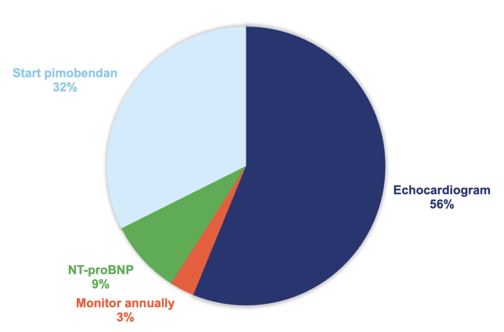
Responding DVMs overwhelmingly selected echocardiography as their next step in this case, highlighting the profession’s continued emphasis on definitive cardiac staging before initiating therapy. While specialists agree that echocardiography remains the gold standard for evaluating myxomatous mitral valve disease, many cardiologists would approach this case differently. Given the breed predisposition, characteristic murmur, and evidence of cardiac enlargement on thoracic radiographs, many specialists would feel there is sufficient evidence to begin pimobendan therapy while pursuing additional diagnostics as needed.
Responding DVMs were asked to identify their next step in the presented case. Over 57% of the responding DVMs selected treating the case by hospitalizing for IV fluids, analgesia, and supportive care. The specialists agree. Read more to learn why.
CLINICAL PERSPECTIVE
How would you handle the case?
A Murmur, Mild Cardiomegaly, and a Clinical Decision
Most cardiologists would agree that this dog almost certainly has myxomatous mitral valve disease (MMVD). The real question is not whether disease is present—but whether sufficient evidence exists to support intervention.
Historically, echocardiography was viewed as the definitive method for distinguishing Stage B1 from Stage B2 disease. However, over the last decade, veterinary cardiologists have invested significant effort in helping general practitioners identify clinically important cardiac enlargement using tools readily available in practice.
Specifically, radiographic measurements such as:
-
Vertebral Heart Size (VHS)
-
Vertebral Left Atrial Size (VLAS)
have become increasingly valuable in helping clinicians determine whether a patient has progressed beyond early-stage disease.
In a breed such as the Cavalier King Charles Spaniel—where MMVD prevalence is exceptionally high—a left apical systolic murmur combined with evidence of cardiac enlargement on thoracic radiographs substantially increases the likelihood that clinically relevant remodeling is already occurring.
For many specialists, this becomes less of a diagnostic question and more of a treatment-timing question.
Why Many Cardiologists Would Start Pimobendan
The EPIC trial fundamentally changed how veterinarians think about preclinical mitral valve disease.
The study demonstrated that dogs with evidence of cardiac enlargement secondary to MMVD experienced a significant delay in the onset of congestive heart failure when treated with pimobendan.
As a result, modern cardiology focuses heavily on identifying dogs that have entered Stage B2 disease before heart failure develops.
In referral settings, echocardiography remains the ideal method for confirming staging. However, in everyday clinical practice, many cardiologists acknowledge that high-quality thoracic radiographs and breed-specific measurements can provide enough information to make informed treatment decisions.
This is particularly true when:
-
The breed is predisposed to MMVD
-
A characteristic murmur is present
-
Radiographic enlargement is already documented
-
Clinical signs of heart failure remain absent
In these cases, delaying treatment solely to obtain an echocardiogram may not always be necessary.
Where Echocardiography Still Fits
From an internal medicine perspective, the most appropriate next recommendation is:
Hospitalize for IV fluids, analgesia, and supportive care
The reason is simple: this dog is already demonstrating evidence of clinically significant disease.
While the laboratory abnormalities are relatively mild, the patient has multiple indicators suggesting systemic involvement:
- Persistent vomiting
- Decreased appetite
- Mild dehydration
- Cranial abdominal pain
- Inflammatory leukogram
- Positive pancreatic lipase screening test
- Breed-associated hyperlipidemia
The combination of these findings suggests a patient that is unlikely to benefit from dietary management alone during the acute phase.
Why Not Simply Monitor?
Monitoring alone may be appropriate for dogs with murmurs but no evidence of cardiac remodeling.
This patient is different.
The combination of:
-
Breed predisposition
-
Characteristic murmur
-
Mild radiographic enlargement
suggests progression beyond the earliest stages of disease.
The concern is not that the dog is currently in heart failure.
The concern is missing the window where intervention has been shown to improve long-term outcomes.
Specialist Takeaway
While echocardiography remains the gold standard for cardiac staging, many contemporary cardiologists would view this case as having sufficient evidence of preclinical MMVD with cardiac remodeling to justify treatment.
For that reason, many specialists would recommend: Start Pimobendan
The broader lesson is that advances in radiographic measurements such as VHS and VLAS have empowered general practitioners to make more informed cardiac treatment decisions without always requiring referral-level imaging.
In this case, the goal is not simply to diagnose heart disease—it is to intervene during the period where therapy has the greatest opportunity to alter the course of disease and improve outcomes.
Clinical Outcomes Annual Report
This report brings together the answers to more than fifteen of this year’s most important clinical questions, offering a comprehensive view of what is working in practices across the country.
Subscribe today and become part of the Clinical Outcomes Network.
Check Out Past Results
See how veterinarians nationwide answered previous Clinical Outcomes questions and what their results reveal.
2 Comments. Leave new
Love the result/pie chart!
Excellent resource